We developed a wide field of view, none-scanning system, to image large regions of retinal explants. Our method allows simultaneous imagine from up to 2 mm2, roughly 20 times larger field of views than done in the past. Although we are currently benchmarking the quality of our recording with experiments gathered by other groups, our preliminary result suggest that we can reproduce previous findings and can use this system in the future to functionally characterize the spatial distribution of retinal responses.
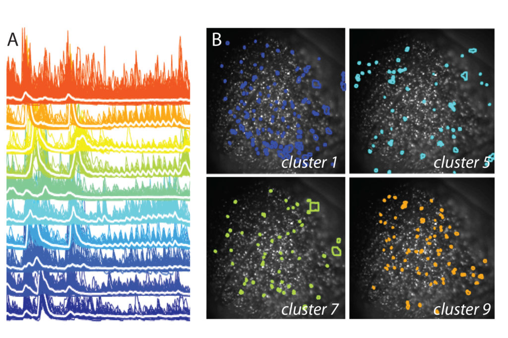
Retinal responses. A. Example traces to full-field flash and chirp stimuli of functionally clustered response properties (colors; white = mean response). B. Retinal images superimposed with the spatial location of particular clustered responses.